
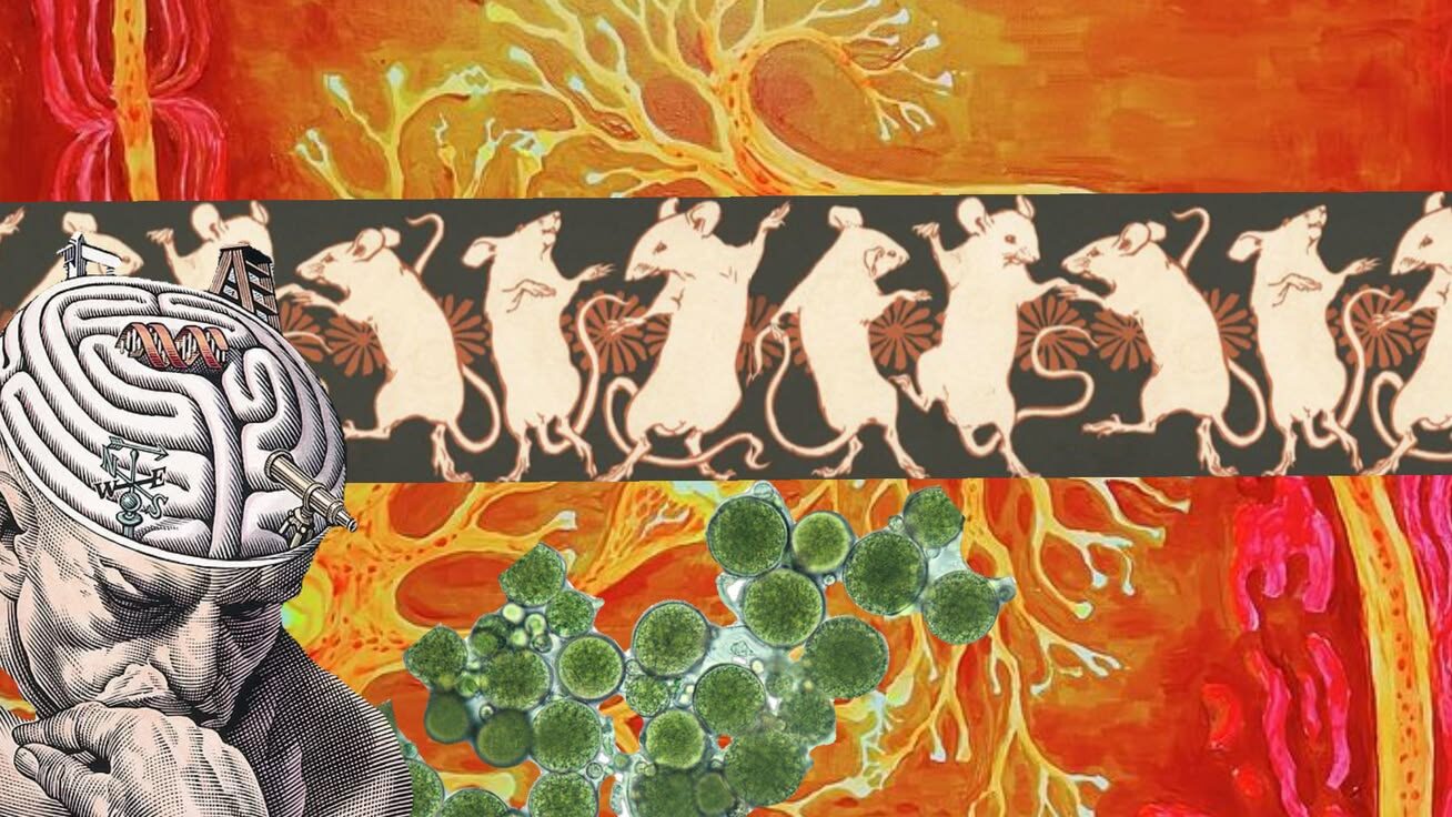

There’s an ongoing debate – primarily among individuals between the ages of 10 and 18 – about which environment would be better to live in: outer space or under water in Earth’s ocean. I would hazard a guess that the most cited evidence for either side would be the comparison of how much we know about outer space compared to the deep sea. Arguments could be made for either, but everyone agrees that we know very little about both of them. Arguably even less well-known is the human brain. Although we understand the cellular biology of the brain quite well, we do not understand very much about how the brain actually works. However, with the emergence of the field of optogenetics, researchers have made significant strides in understanding the brain. Breakthroughs have been made in regards to memory and emotions and mental illness as well as in clinical applications to neurodegenerative disorders like Parkinson’s, Alzheimer’s, and retinitis pigmentosa. Recently, the Nobel Prize in Physiology or Medicine was awarded for optogenetics to Karl Deisseroth, Georg Nagel, and Peter Hegemann for their contributions to the field.
Optogenetics is a relatively new field in neuroscience that takes advantage of light responsive proteins to exert extremely precise control over events in biological systems. One of the pioneers in the field Karl Diesseroth stated that, “By delivering optical control at the speed (millisecond-scale) and with the precision (cell type–specific) required for biological processing, optogenetic approaches have opened new landscapes for the study of biology, both in health and disease.” (Deisseroth, 2010).
Although the Nobel prize for optogenetics was awarded this year, the study of light gated proteins goes back to 1971 when Dieter Oseterhelt and Walther Stoeckenius discovered the light-driven ion pump Bacteriorhodopsin in the plasma membrane of the halophilic archaeon Halobacterium halobium. The path to a breakthrough was slow and steady from that point on. Two years later, they showed that Bacteriorhodopsin was an outward proton pump capable of hyperpolarizing the cell when stimulated by visible light. In 1977, Akemi Matsuno-Yagi and Yasuo Mukohata identified a second light-driven ion pump in H. halobium when they were working with a bacteriorhodopsin-deficient mutant strain that hyperpolarized when exposed to light. This second pump is known as Halorhodopsin and hyperpolarizes cells by pumping chloride ions into the cell when stimulated by light.
In the 1990s, the investigation into light-driven ion pumps gave way to a discovery that would truly enable the field of optogenetic when Peter Hegemann discovered channelrhodopsin in the unicellular green algae Chlamydomonas reinhardtii. When the organism was stimulated with light, he found that “the phototactic behavior was extraordinarily fast, with a latency of only microseconds after flash stimulation” (El Manira et al., 2026). The response was too fast to involve typical phototransduction cascades. This observation led to the crucial idea that the rhodopsin photoreceptor and the ion pumps must be linked. The direct connection between rhodopsin photoreceptor and ion pumps was the gateway to being able to isolate and control cells in biological systems with a temporal precision greater than ever before.
Soon after, the first true big breakthrough in Optogenetics came in 2002 when Georg Nagel, Peter Hegemann, and Ernst Bamberg expressed the Chop1 mRNA encoding in frog eggs. When the oocytes were exposed to green-light illumination, the cell was flooded by protons and depolarized. The speed at which the cell depolarized led to the first evidence of a light-gated ion channel and the distinction of Channelrhodopsin-1. This was significant because ion channels can carry ions across the cell membrane much faster than ion pumps since channels facilitate passive transport. The next year, Nagel, Hegemann, and Bamberg made the functional distinction between Channelrhodopsin-1 (ChR1) and Channelrhodopsin-2 (ChR2). While ChR1 is activated by green light, ChR2 is activated by blue light.
As cool as making algae and frog eggs move with lasers, scientists were still looking for a way to apply this technology to the human brain. At the time, one of the primary challenges in the field of neuroscience was the inability to isolate control in specific cells in the brain with the necessary spatial and temporal precision. Even though bacteriorhodopsin and halorhodopsin had been identified, there was no connection between light activated proteins and human neurons. It wasn’t until 2005 that Karl Deisseroth showed that ChR2 could be expressed in neurons. Using mammalian neurons transduced with ChR2, he demonstrated “reliable, millisecond-timescale control of neuronal spiking, as well as control of excitatory and inhibitory synaptic transmission” (Boyden, 2005). This showed just how vast the applications of optogenetics could be. Researchers now had a way to control action potentials in mammalian neurons with the speed and localization they needed to adequately study a human brain.
The field of optogenetics has spread to include not only studies of the brain and other biological systems but also clinical applications to neurodegenerative diseases. Among these applications, efforts have been made in treatments for Parkinson’s and retinitis pigmentosa.
Understanding the Human Brain
At this point, the primary focuses of optogenetically driven studies of the brain have focused on mapping and manipulating connections in the brain. Before, scientists could only observe brain activity and look for patterns. They could correlate activities with behaviors but had no way to prove the actual causes. One of the most crucial advancements made via optogenetics is the newfound ability to create brain activity rather than merely observing it. By making specific neurons respond to light, researchers can now directly activate or deactivate neurons and observe the effects of their manipulations.With this ability, researchers have been able to map brain circuits to behavior, emotions, and cognition (El Manira et al., 2026).
A pair of particularly interesting studies on memory showed that these methods could be used to induce memory recall and even create artificial memories in mice. Researchers exposed the mice to contextual fear conditioning while expressing ChR2 in certain neurons. The mice were returned to a neutral environment, but when their targeted neurons were optically stimulated they exhibited the same fear response (Liu, et al., 2012). In a separate experiment they stimulated neurons that were naturally active in the neutral context during fear conditioning. When the mice returned to the neutral context that time, they exhibited the same fear response despite being in the neutral context. The mice had never been fear conditioned in the neutral environment yet they showed the fear response anyway, indicating an artificially generated memory (Ramirez, et al., 2013).
Applications: Mental health/emotions
Optogenetics has been used to prove that it is not specific regions and units of the brain that control emotions but rather very specific, discrete circuits. This has changed how scientists think about mental illness. Karl Deisseroth claimed that the inability to probe precise neural circuits is why our understanding of brain disorders is so underdeveloped since the brain is itself an extremely complex and high speed neural circuit. He said, “rather than conceptualizing the brain as a mix of neurotransmitters, ideally we would be able to move toward a circuit-engineering approach, in which devastating symptoms of disease are understood to causally result from specific spatiotemporal patterns of aberrant circuit activity relating to specific neuronal populations” (Deisseroth, 2010).
Applications: Vision
One of the clinical applications of optogenetics could be the restoration of vision after being affected by neurodegenerative conditions like Retinitis pigmentosa, which is a hereditary disease that leads to the loss of rods and cones in the eyes eventually resulting in blindness. In a 2006 study, researchers injected ChR2 into the eyeball of mice and expressed ChR2 in cells that connect the retina to the brain via the optic nerve. With light stimulation, the cells depolarized and retinal photosensitivity was restored allowing light signals to reach the visual cortex. This study proved that expressing ChR2 in any remaining retinal neurons could be a strategy for vision restoration in cases of retinitis pigmentosa in cases of incomplete photoreceptor degeneration (Bi, et al., 2006).
Applications: Parkinson’s
Two prominent applications of optogenetics to Parkinson’s Disease are identifying its pathogenesis and providing a treatment therapy. We know a lot about how Parkinson’s Disease affects people, but the root cause is still unknown. Researchers are hoping to use optogenetics to investigate the pathogenesis of Parkinson’s. It is speculated that the ability to target specific cell types for manipulation via optogenetics will prove to be a very accurate method for discovering more about the origin of the disease (Bi, et al., 2025).
Although the cause of Parkinson’s is still unknown to us, researchers have already begun testing optogenetics as a potential treatment method for the disease. The current treatment can involve medication that has some very undesirable side effects and/or highly selective and invasive surgery (Li et al., 2026). Some researchers have proposed a specific type of minimally invasive optogenetics called upconversion optogenetics as a potential treatment for Parkinson’s. They have already done studies on mice with Parkinson’s using this method that have shown it can cause improvement in coordination as well as overall motor function (Li et al., 2026).
Beyond the potential to just treat Parkinson’s Disease, this method of upconversion optogenetics is also significantly less invasive than conventional methods which “still face the disadvantage of high invasiveness due to the poor penetration of short-wavelength visible light in brain tissue. This necessitates the delivery of light through inserted optical fibers, particularly in deep brain regions, resulting in severe brain damage, acute inflammation, and long-term immune responses.” (Li et al., 2026).
TLDR:
Optogenetics is a field that uses light responsive proteins to control events in biological systems to study biology and neuroscience. It has provided a way to precisely exert control over neurons (and other targeted cell types) that has proven crucial to the study of how the human brain truly works. This year, the Nobel Prize in Physiology or Medicine was awarded to three researchers who are pioneers in the field. Relatively new, the field has been progressing since the 1970s.
Simplified Discovery Timeline:
1971 – First light-driven ion pump, bacteriorhodopsin, discovered in the plasma membrane of Halobacterium halobium.
1973 – Showed that bacteriorhodopsin was an outward proton pump that could hyperpolarize cells upon light stimulation.
1977 – A second light-driven ion pump, halorhodopsin, is identified in H. halobium. It hyperpolarizes cells by pumping chloride ions into the cell when stimulated by light.
1990s – The direct connection between the rhodopsin photoreceptor and ion pumps is established based on the speed of response to light stimulation.
2002 – First evidence of light-gated ion channels and distinction of Channelrhodopsin-1 as a light-gated ion channel.
2003 – Functional distinction made between green light sensitive Channelrhodopsin-1 and blue light sensitive Channelrhodopsin-2.
2005 – Channelrhodopsin-2 expressed in mammalian neurons in vivo.
2010 – Channelrhodopsin, bacteriorhodopsin and halorhodopsin all capable of quickly turning neurons on or off safely.
The field of optogenetics has spread to include not only studies of the brain and other biological systems but also clinical applications to neurodegenerative diseases. Among these applications, efforts have been made in treatments for several neurodegenerative diseases.
Citations:
Bi A, Cui J, Ma YP, Olshevskaya E, Pu M, Dizhoor AM, Pan ZH (2006) Ectopic expression of a microbial-type rhodopsin restores visual responses in mice with photoreceptor degeneration. Neuron 50:23-33
Bi, Y., Wang, Y., Gong, L., Ren, Y., & Guo, Y. (2025). Insight into the application of optogenetics for regulating motor-related circuits in parkinson’s disease and novel therapeutic target exploration. Parkinsonism & Related Disorders, 139, 107988. https://doi.org/10.1016/j.parkreldis.2025.107988
Boyden, E. S., Zhang, F., Bamberg, E., Nagel, G., & Deisseroth, K. (2005). Millisecond-timescale, genetically targeted optical control of neural activity. Nature neuroscience, 8(9), 1263–1268. https://doi.org/10.1038/nn1525
Deisseroth, K. (2010). Optogenetics. Nature Methods, 8(1), 26–29. https://doi.org/10.1038/nmeth.f.324
El Manira, A., Svenningsson, P., & Linnarsson, S. (2026, October 5). Scientific background 2026 Optogenetics Discovery of a Neuronal Switch. https://www.nobelprize.org/uploads/2026/10/advanced-medicineprize2026.pdf
Li, X., Wang, X., Zhou, T., Yu, J., Xiang, H., Zhang, C., Sun, S.-K., & Liu, R. (2026). Minimally invasive upconversion optogenetics for Parkinson’s Disease Treatment. Biomaterials, 324, 123509. https://doi.org/10.1016/j.biomaterials.2025.123509
Liu X, Ramirez S, Pang PT, Puryear CB, Govindarajan A, Deisseroth K, Tonegawa S (2012) Optogenetic stimulation of a hippocampal engram activates fear memory recall. Nature 484:381-385.
Ramirez S, Liu X, Lin P-A, Suh J, Pignatelli M, Redondo RL, Ryan TJ, Tonegawa S (2013) Creating a false memory in the hippocampus. Science 341:387-391.
