
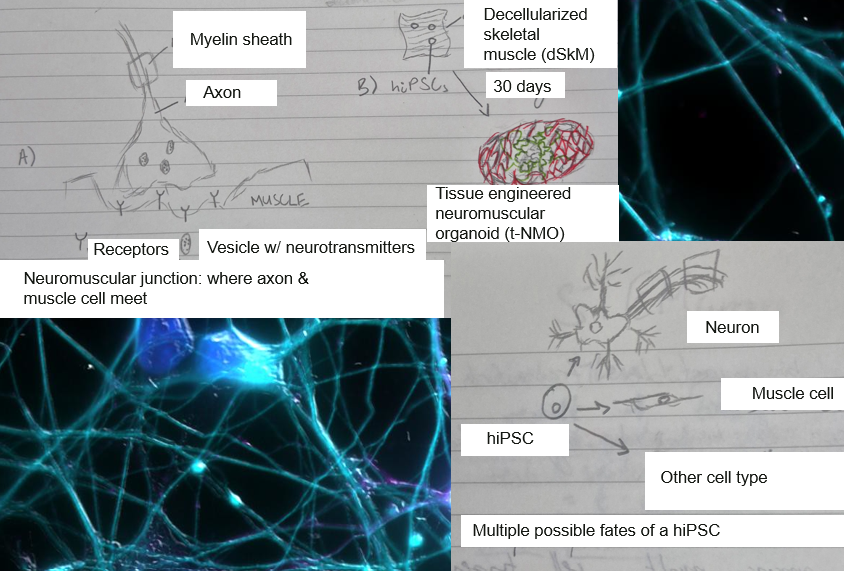

While scrolling through Instagram, I came across a researcher who worked with “organoids”. The futuristic name was enough to draw me in. I found that, as the name implies, these structures are organ-like. Their (intended) purpose is to mimic the environment and events that occur naturally in the organized structure of an organ. Scientists are able to study basic biology and generate disease models by recreating these characteristic, naturally occurring interactions outside of the living system.
Tissue Engineered Neuromuscular Organoids (t-NMOs)
In the summer of 2025, researchers Beatrice Auletta and Pietro Chiolerio announced the latest breakthrough in the field of tissue engineering. They had created a tissue-engineered neuromuscular organoid (t-NMO) that reflected the physiological behaviors of motor neurons and skeletal muscle cells. Two essential ingredients are required to create a t-NMO. The first is human induced pluripotent stem cells (hiPSCs). These cells have the ability to differentiate (transform from an unspecialized cell to a specialized cell with a unique function). The second ingredient is what hiPSCs grow on. Compared with differentiating hiPSCs in flasks like others before them (Faustino Martins, 2020), the researchers were able to differentiate hiPSCs in the biological scaffold of decellularized skeletal muscle (dSkM). The biological scaffold provides structure and a biochemical network. Here, the researchers stripped donor muscle tissue of living cells, cellular debris, DNA, and RNA. The only thing left was the extracellular matrix (ECM): a 3D structure consisting of structural and functional macromolecules. In this context, dSkM acts as the foundation of a house while hiPSCs serve as the builders. The finished house is the t-NMO.
Flask or Scaffold?
Auletta and Chiolerio also cut the amount of time needed to grow the organoid from 40 days to 30 days, saving scientists time and resources (Auletta, 2025). The differences in differentiation environment (flask versus biological scaffold) influenced the way the cells grew, promoting continuity of growth compared with choppier self-assembly. This study supported the claim that differentiation of cells on a natural ECM scaffold encourages natural connections and behaviors between different cell types. In contrast, differentiation and growth without this natural scaffold is less representative of actual growth conditions, producing interactions between cell types that are more strained.
Basic Biology
Auletta and colleagues have proven that an extracellular matrix scaffold can be used to produce more natural cell-cell interactions that mimic organ spaces. This application is especially helpful in advancing our understanding of basic biology. By studying this new system, scientists are able to learn more about the driving mechanisms behind activity at the neuromuscular junction (NMJ). An NMJ refers to the synapse between a neuron and a muscle cell. These synapses are studied because they are easier to access compared to synapses located deep inside the nervous system.
Defects in NMJs lead to serious conditions such as Duchenne Muscular Dystrophy (DMD). DMD is a genetic disorder that results in heart and skeletal muscle weakness. The condition worsens quickly over time, and there is currently no cure (Cleveland Clinic). Because DMD is the most common form of muscular dystrophy, researchers are pushing to better understand the disease mechanism and improve current treatment.
Disease Models
The organoids created by the researchers display phenotypes characteristic of healthy neuromuscular function. The scientists also created a disease model using their new protocol. Instead of using hiPSCs from a healthy donor, they seeded dSkM with hiPSCs from a patient afflicted with DMD. The resulting t-NMO displayed phenotypes characteristic of DMD. Directly comparing these healthy and diseased organoids helps researchers pinpoint exact cellular and molecular differences in a human-like environment.
Conclusions
Organoids are a scientific tool that allow researchers to closely study relevant diseases and inform the design of future treatments. Advancements in organoids translate to more accurate analysis of disease models, with the ultimate goal of designing better treatments.
References
Auletta, B., Chiolerio, P., Cecconi, G., Rossi, L., Sartore, L., Cecchinato, F., Barbato, G., Lauroja, A., Maghin, E., Easler, M., Raffa, P., Angiolillo, S., Qin, W., Frison, R., Calabrò, S., Villa, C., Gagliano, O., Laterza, C., Cacchiarelli, D., … Urciuolo, A. (2025). Tissue-engineered neuromuscular organoids. Communications Biology, 8(1). https://doi.org/10.1038/s42003-025-08484-z
Cleveland Clinic. (2022, July 25). Duchenne muscular dystrophy (DMD): Symptoms & treatment. Cleveland Clinic; Cleveland Clinic. https://my.clevelandclinic.org/health/diseases/23538-duchenne-muscular-dystrophy-dmd
Faustino Martins, J.-M., Fischer, C., Urzi, A., Vidal, R., Kunz, S., Ruffault, P.-L., Kabuss, L., Hube, I., Gazzerro, E., Birchmeier, C., Spuler, S., Sauer, S., & Gouti, M. (2020). Self-organizing 3D human trunk neuromuscular organoids. Cell Stem Cell. https://doi.org/10.1016/j.stem.2019.12.007
